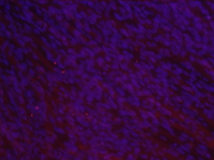
Immunofluorescence of Muscle Stem Cells After Development
I studied the growth of C2C12 myoblast cells with Dr. Balza. I focused on learning aseptic and immunofluorescence techniques, and animal cell incubation.
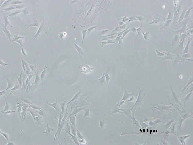
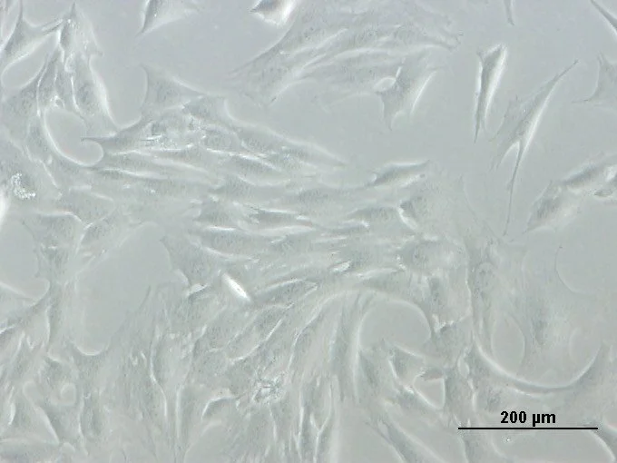
I observed the confluence of a sheet of cells by observing the cells through a microscope daily. These are some images of cells I gathered after 24 hours at about 20% confluence.
Once the cells were at 80% confluence, I used immunofluorescence to visualize the myosin fibers and nuclei. They were the edited together using photoshop.